Over the last 60 years, the primary imaging of the central lymphatic ducts was performed using a pedal lymphangiogram. Traditional pedal lymphangiography is both time-consuming and technically challenging and requires specialized not readily available equipment. The recent development of US-guided intranodal lymphangiogram significantly simplified the imaging of the central lymphatic system and opened the door to a wider spread of lymphatic interventions from specialized academic centers to community hospitals1,2. Based on a simple US-guided puncture of the groin lymph nodes this procedure is within basic skills of any interventionalist practicing US-guided procedures. During the last few years, intranodal lymphangiogram has almost completely replaced the traditional pedal lymphangiogram due to its simplicity, high rate of success, and equipment availability.
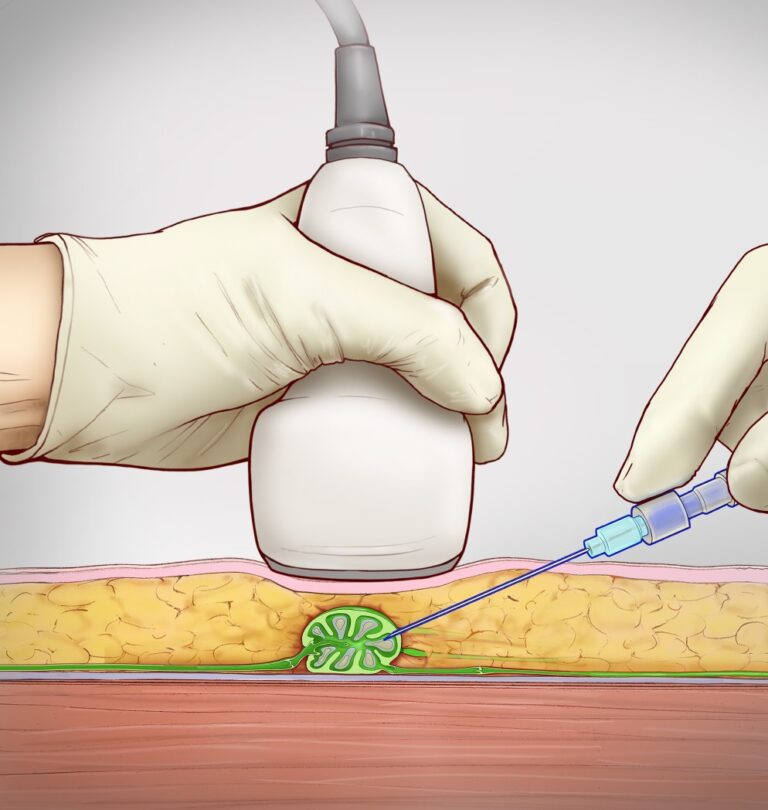
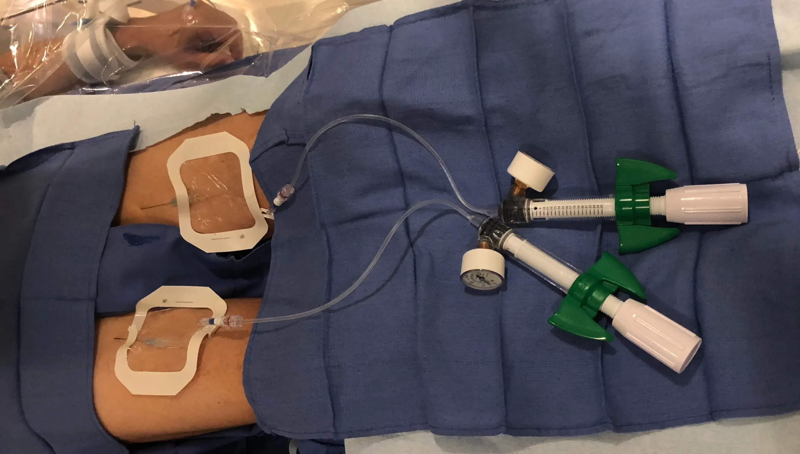
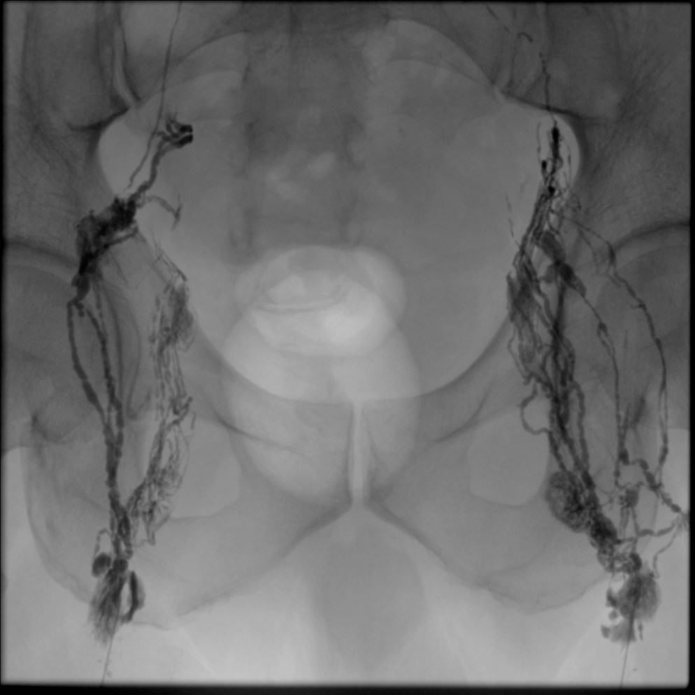
In this technique, an inguinal lymph node is directly accessed under ultrasound guidance with a 25-gauge spinal needle with the needle tip positioned in the hilum of the node (Figure 1). Subsequently, an oil-based contrast agent (Lipiodol, Guerbet Group, Princeton, NJ) is injected at a rate of about 1-2 mL per 5 minutes (Figure 2). If successful, immediate opacification of the lymphatic vessels is observed under fluoroscopy (Figure 3).