Even though the lymphatic system is known for hundreds of years, its imaging was practically non-existing until recently. Lack of imaging lead to lack of our understanding of the some of the important pathophysiological mechanisms.
One of the additional obstacles is that the lymphatic system consists of several subsystems that communicate with each other in a very complicated way. In a sense the lymphatic system can be presented as a reversed tree, where the thoracic duct (the main aqueduct of the lymphatic system) is a trunk and all other lymphatic system drain into it.
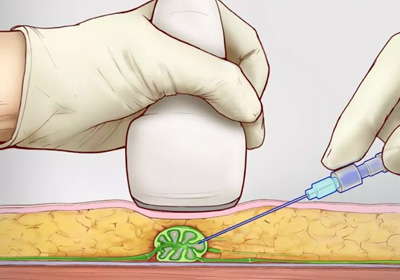
The most important lymphatic system in the body are liver, intestine and soft tissue. In normal patient, the flow in each system is unidirectional toward TD. In order to opacify these systems, the dye has to be introduced in each system separately (Figure).
For opacification of the soft tissue systems, the contrast is introduced through the groin lymph nodes (intranodal lymphangiography or DCMRL).
For opacification of the liver lymphatic system, we perform liver lymphangiography.
For opacification of the mesenteric lymphangiography, we perform open or percutaneous mesenteric lymphangiography.