Dynamic Contrast Enhanced MR Lymphangiography (DCMRL)
About DCMRL
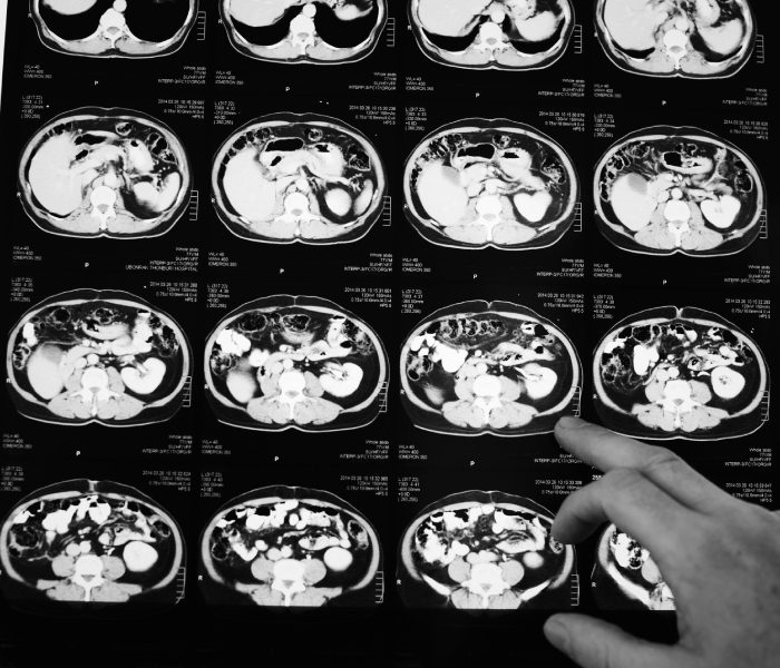
Magnetic resonance provides outstanding tissue resolution and especially sensitive for the presence water. It is obvious, that evaluation of the lymphatic system would be of incredible value.
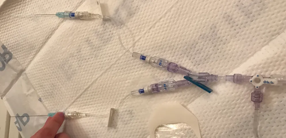
DCMRL was developed approximately five years ago in order to image the central lymphatic system in a way similar to the intranodal lymphangiography1,2. It is performed by placing of the small needles into the groin lymph nodes. It is usually performed outside the room where is the MR magnet is located. It is much easier to do when MR equipped with detachable table. The table detached, moved in the prep area and the needle are inserted.
To confirm the position of the needles we usually inject the US contrast (Sonovue, Bracco Monroe Township, NJ) through the needles. The proper position is confirmed, when the draining lymphatics ducts are enhanced3(Movie).
The patient is then positioned in the MR scanner and gadolinium-based contrast agent (Dotarem, Guerbet Group, Princeton, NJ) is injected into the lymph nodes.The acquisition techniques include three elements (we using Siemens equipment, however, all other manufactures have similar sequences):
01. Water imaging (HASTE, Heavy TW2 sequences)
- 1. Lymphatic masses (Figure)
- 2. Effusions (fluid in the cavities)
- 3.Edema
02. Fast angio acquisition technique (TWIST)
- 1. Dynamic evaluation of the lymphatic flow (how fast lymph flows)
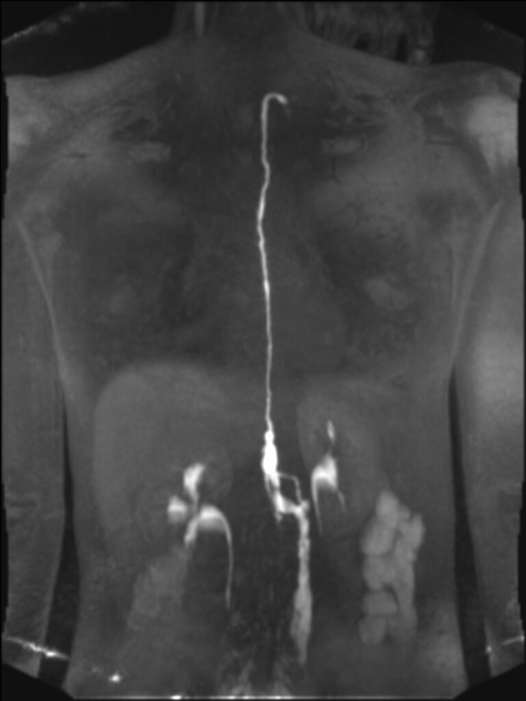
03. Delayed high resolution angio technique (Inversion recovery with fat suppression)
- 1. Allows for delayed images, when the timing of the contrast material is slower
- 2. High resolution images of the lymphatic system
What lymphatic pathology DCMRL can show?
- Chylothorax
- Plastic bronchitis
- Chyloptysis
- Interstitial lung disease